Medical imaging, made visual.
Short animated lessons on radiology, interventional radiology and imaging for every specialty.

Can you spot it?
Tap where this head CT looks different.
Unenhanced head CT. Compare the two sides, then tap the area that looks abnormal.
What Contrast is
Real scans, live contours
Every lesson is built on real, openly licensed scans, with the finding outlined in gold as you watch.
Animated lessons & IR procedures
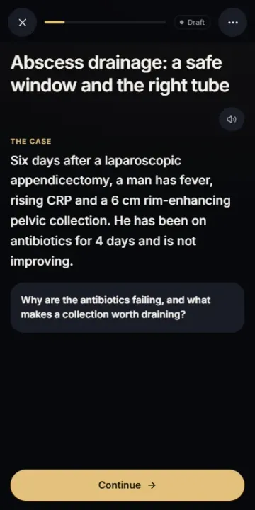
Signs, artefacts and imaging physics, and interventional procedures step by step, in a minute or less.
Spot it: learn by playing
Find the finding before the answer. Short image puzzles that train your eye.
The Contrast app
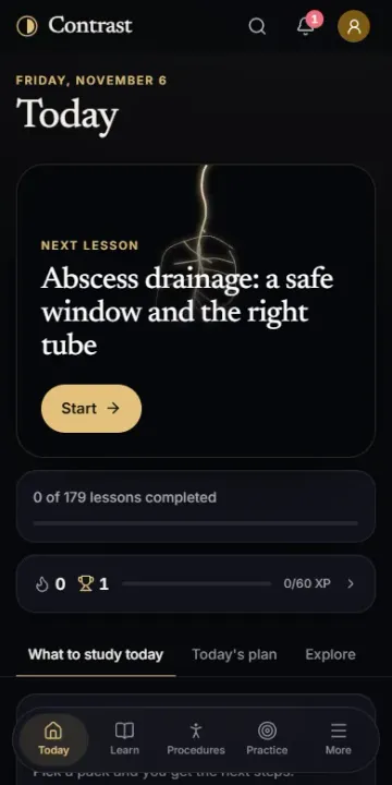
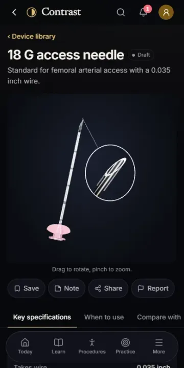
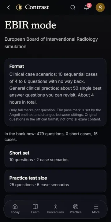
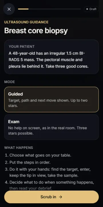
Signs atlas, image cases, IR devices in 3D, procedure simulations and mock exams, in English and Turkish.
Latest lessons
All lessons →
CT windows: one scan, many pictures
Why the same CT slice can look completely different: Hounsfield units, the limited greys of a screen, and why scans are read in more than one window.

Early ischaemic signs on unenhanced CT
The subtle signs of an early middle cerebral artery infarct on a plain head CT: the insular ribbon, the lentiform nucleus and the grey-white border.
Pulmonary embolism on CT: the filling defect
How a pulmonary embolus looks on CT pulmonary angiography, how acute and chronic clot differ, and why a poor study should say what it could not assess.
Be first to try Contrast
Join the waitlist: we will e-mail you when the app opens, plus a short newsletter with new lessons. Unsubscribe any time.
Follow along
One finding a week on YouTube, Instagram, TikTok and X.